Delayed In Vitro Fertilization Using Coho Salmon Ovarian Fluid
(Source: G.E. Corley‑Smith, C.J. Lim, and B.P. Brandhorst from Zebrafish Book 5th Edition)
Overview of technique
For successful in vitro fertilization, zebrafish eggs must normally be fertilized almost immediately after being collected from the female. However, zebrafish eggs can be held in Coho salmon (Oncorhynchus kisutch) ovarian fluid for periods exceeding 1.5 hours with high subsequent fertilization rates. Eggs can be successfully held for periods exceeding 6 hours, although the fertilization rate is compromised. For a general discussion of in vitrofertilization, please see *Embryo Production By In Vitro Fertilization (page 2.14).
To delay fertilization of eggs after they are extruded from the female zebrafish, eggs are held at room temperature (18‑22°C) in the ovarian fluid (the fluid that surrounds mature eggs) of Coho salmon. In collecting ovarian fluid, contamination by broken salmon eggs, blood and water should be minimized. The female salmonid from which ovarian fluid is collected should be mature and ready to deliver eggs but should not be overly ripe with broken eggs and watery ovarian fluid. Ovarian fluid from a non‑ripe females may be useable, but the volume of ovarian fluid from a non‑ripe female is smaller. We collect ovarian fluid at a salmon hatchery.
As batches of ovarian fluid vary, we test individual batches of ovarian fluid. When a batch (ovarian fluid collected from one fish) is identified that allows for holding zebrafish eggs for 1.5 hours with high subsequent fertilization rates, we aliquot this batch into 1.5 ml screw cap microcentrifuge tubes and store frozen at ‑20°C or ‑80°C. Coho salmon ovarian fluid is robust and can undergo repeated freezing and thawing and can be left for several days at room temperature and still function. However, for optimal fertilization rates we suggest more careful storage.
High fertilization rates can only be achieved when good quality zebrafish eggs are obtained. It is possible that females produce and resorb eggs on a daily cycle and eggs may be in peak condition for fertilization for only a short period. Some good zebrafish facilities routinely collect eggs in the first few hours after the fish are exposed to the light phase of the photocycle. However, some of our best eggs are collected later in the day. When we desire high fertilization rates, we perform the following time consuming procedure. Shortly after the beginning of the light part of her photocycle, the female is placed in a small tank with at least one male. The fish are observed and as soon as breeding activity commences the female is removed from the tank and squeezed for eggs. Good eggs are slightly granular and yellowish in color. Although not always completely spherical, the best eggs look full. The best batches contain few, if any, broken eggs and no whitish or withered eggs. The chorion of good eggs can be observed under a stereoscopic dissecting microscope to elevate up away from the plasma membrane when ovarian fluid is diluted with water. An elevated chorion indicates that the egg is no longer capable of being fertilized. Thus, we often test a few eggs from each female by adding water to them. Some of the batches of milt we view under the microscope include many sperm that have a short knobby tail. These sperm are usually poor swimmers and are suspected to give poor fertilization rates. We do not use batches of milt containing such sperm.
Sperm held in sperm extender (recipe is provided at end of this article) at 4°C for up to 8 hours are still capable of a low rate of fertilization. Thus, it is possible to collect sperm at the same time eggs are collected; however, we have observed that fresh sperm yields higher fertilization rates. Sperm can usually be collected at any time of day. When milt is diluted with water, the sperm become motile for about 1 minute, during which time they can fertilize eggs. The capacity of sperm to be activated with water can be monitored. Place a few μl of sperm in extender on a microscope slide and place a cover slip over it. Use phase contrast or DIC under high power to visualize sperm. Add a few drops of water along the edge of the cover slip, and observe if the sperm actively swim as the water streams under the coverslip.
The order in which eggs and sperm are collected will vary depending on the experiment. Eggs and sperm are collected initially into glass microcapillary tubes. Gentle suction can be applied by several methods, including mouth pipetting.
Collection of Coho Salmon Ovarian Fluid:
Coho salmon are terminal spawners; thus, eggs are normally collected at hatcheries immediately following lethal cranial trauma. The euthanized female Coho salmon is immediately hung by its tail and all the gills on both sides of head are slit to drain blood. Pushing a wad of paper towels under the operculum will help soak up blood and impede coagulation. After 5 minutes, dry the fish with a towel so that no water can drip into eggs and ovarian fluid when they are collected. Have an assistant hold the fish by its head and tail, with its belly downward over a clean dry bowl, and slit the fish from anus to the front of the body cavity. Eggs and ovarian fluid will fall into bowl. Release eggs from skein (ovarian connective tissue) and remove pieces of skein from bowl containing free eggs and ovarian fluid. Remove approximately 75% of ovarian fluid to 50 ml conical plastic tube. A non‑abrasive kitchen strainer or colander, stainless steel or plastic, is useful for separating eggs from ovarian fluid. Handle eggs gently at all times to prevent breakage. Give the eggs to hatchery staff and put the ovarian fluid on ice. Back at the lab, centrifuge the tubes of ovarian fluid at 5500g for 5 minutes at 4°C to sediment cellular debris. Remove the supernatant and freeze.
Collection of Zebrafish Eggs for Delayed In Vitro Fertilization
1. Place approx. 100 μl salmon ovarian fluid in 50mm diameter Petri dish at room temperature. It should form a small dome near the center of the dish.
2. Anaesthetize a female zebrafish.
3. Place female belly up under dissecting scope, resting in a V‑shaped slit of a damp sponge.
4. Carefully dry belly and genital pore with Kleenex® or Kimwipe®.
5. Draw some of the ovarian fluid from the Petri dish up in a silanized glass capillary tube (Kimax‑51, Kimble Products Art. No. 34502, ID 0.8‑1.1mm, length 100mm) and expel back into the dish. This is done to pre‑wet the tube, reducing friction and lessening the chance of breaking eggs.
6. Squeeze fish gently and gently draw eggs up into the glass capillary tube.
7. Observe eggs under dissecting scope and assess quality by appearance.
8. Gently expel eggs into ovarian fluid on the Petri dish. Try to avoid placing eggs on dry dish, which may reduce subsequent fertilization rates.
9. Return fish to water.
10. Place lid on Petri dish to reduce evaporation. Although perhaps not necessary, we place black plastic sheets over the dishes to shield out light.
11. Eggs are held at room temperature until fertilization is desired.
Collection of Sperm for Delayed In Vitro Fertilization
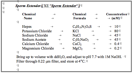
1. Put 50μl sperm extender (see Sperm Extender, page 7.36) into 500μl microcentrifuge tubes in ice.
2. Perform steps 2 through 4 as described above for female.
3. Squeeze fish gently and take up milt into a 2μl or 5μl glass capillary tube (Drummond Microcaps) by capillary action. This tube does not need to be silanized.
4. Gently expel milt from one male into microfuge tube containing sperm extender that is on ice: typically 1‑2μl/50μl sperm extender.
5. Gently swirl to mix sperm and sperm extender.
6. Store on ice until needed.
In Vitro Fertilization
1. First, remove most of the ovarian fluid from the eggs with a sterile pipette to a clean 0.5 ml microcentrifuge tube. Later when time permits, centrifuge at 5500g for 5 minutes at 4°C, decant supernatant and freeze. Ovarian fluid can be reused several times.
2. Spread 5‑15 μl of sperm extender containing sperm evenly over all the eggs in the Petri dish.
- Immediately add 0.5 ml fish water and swirl very gently to mix.
- After 1 minute, very gently add 28.5°C water to 3/4 fill Petri dish and leave at 28.5°C for 1 hour. To promote gas exchange, make sure there is a space between top of water and Petri dish cover.
5. After 1 hpf, add 0.02% stock solution of methylene blue for a final concentration of 0.3 PPM. This helps to inhibit fungal growth. Make sure eggs do not touch each other, to reduce the spread of fungus that may grow on dead eggs from attacking developing embryos.
6. Place in 28.5°C incubator.
7. After 24 hpf, remove dead embryos and flush out methylene blue.
Sperm Extender: